Growing Pains
An issue we see regularly at Foot & Leg Pain Clinics
Clinics located across Melbourne. Click here for locations
Introduction
Any Sports Podiatrist working with young active kids will know that they are at risk of overuse injuries due to their immature musculoskeletal systems. However, it is imperative that practitioners can confidently identify when children require a professional intervention rather than dismissing their symptoms just as ‘growing pains’.
It has been found that a high proportion of all injuries in sport are due to overuse, and young kids, especially early teenagers tend to partake in high volumes of physical activity. These children can potentially be playing sport at clubs and national levels as well as participating in physical education at school and other organised sports.
It should be not be assumed that children are miniature adults and it should be noted that some of their injuries are unique to the growing years. So when injuries are similar we should still consider the additional effects of growth spurts on these structures and this is when challenges can occur.
Background Information with "growing pains" of the knee
Osgood-Schlatters is a traction apophysitis that is most common in male athletes aged 13-14 years old, and can be bilateral in up to 50% of cases. Girls tend to be affected aged 10-11 years old. Unfortunately the exact incidence and prevalence of the condition is unknown.
Anatomy
Although the general anatomy (eg ligaments, muscles) of the knee is the same in children and adults there are some significant differences related to the ‘growth plates’ that makes children prone to bony injuries rather than ligamentous or muscular damage. This is also the cse with growing feet. legs and hips.
Apophysis
- Where the musculotendinous unit inserts
- Vulnerable to traction injuries as muscle and tendon growth may be slow relative to bone growth
Epiphysis
- The actual site of bony growth
- Inherently unstable and vulnerable to shear forces which can damage bone structure through slips or fractures
Osgood-Schlatters, specifically can be described as an apophysitis, as the symptoms occur where the quadriceps attaches to the tibial tuberosity of the knee. As traction is applied to the tibial tuberosity the apophysis of the tuberosity eventually separates from the tibia. After initial fragmentation at the site, ossification eventually leads to an increase in bone. Thus giving the characteristic prominent tibial ‘bump’.
Subjective Assessment
The child suffering from Osgood-Schlatters will often report:
- Mechanism: kids often report a gradual onset in anterior knee pain but often symptoms will be exacerbated by pubertal growth spurts.
- Pain Area: the pain is often localised around the tibial tuberosity and patellar tendon. However, be aware that in more severe cases, pain can be distributed rather diffusely if repetitive stress is placed on an immature patellar tendon insertion.
- Aggs: Running, Jumping, Kneeling. Descending stairs
- Eases: Rest, Activity modification, Orthotic intervention
Objective Assessment
Diagnosis for this condition tends to be clinical and quite simple. Therefore, much of the objective examination is aimed at identifying potential musculoskeletal and biomechanical contributors to pathology that are amenable to treatment. You will often find:
- A prominent, tender tibial tuberosity
- Quadriceps usually tight
- Ankle Dorsiflexion can be reduced, and is often associated with genetic bony ankle joint shape.
- There are often other biomechanical contributors present, including excessive pronation, tibial or femoral internal rotation
Differential Diagnoses
There are a few differential diagnoses that you should be aware when assessing the young athlete with anterior knee pain, these include:
- Patellofemoral pain syndrome
- Patellar tendinopathy/tendon injury
- Sinding-Larsen-Johansson syndrome
- Osteochondritis Dissecans
- Bursitis
- Fat Pad Impingement
- Referral from hip: including Slipped Capital Femoral Epiphysis and Perthes Disease
- Red Flag conditions – infection, malignancy
Imaging
The use of imaging is not required routinely with this client group as the diagnosis can be made quite simply via a thorough clinical examination. However is symptoms are severe and unrelenting further investigations are merited to rule out other more serious conditions (eg malignancy, infection). Imaging should also be utilised if there are any doubts surrounding a diagnosis of osteochondritis dissecans, slipped capital femoral epiphysis or Perthes Disease.
Standard radiographs can confirm Osgood-Schlatters by revealing heterotropic ossification at the site of the tubercle.
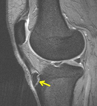
MRI findings will vary dependant on the stage of the condition but can highlight additional soft tissue changes that can occur. These include soft tissue swelling anterior to the tibial tuberosity, loss of the sharp angle to the infrapatellar fat pad, thickening and oedema of the patellar tendon and infrapatellar bursa (2).
Management of Osgood Schlatters Disease
Conservative management involves a significant amount of education of the child, parents and even coaching staff, as their co-operation is often required during the implementation of the childs rehabilitation.
- Poor foot posture and joint movement issues need to be addressed, consider these options for controlling excessive pronation. Regular icing of the knee post activity for pain relief.
- Correcting biomechanical contributors is essential. This may include tightness and weaknesses present in surrounding musculature. For tightnesses consider the calf, hamstring and quadriceps. Regarding weaknesses assess the pelvic stabilisers, medial quadriceps, extrinsic and intrinsic foot musculature.
- Painkillers/NSAIDs should be avoided, although may be indicated around competition periods as required.
- Stretching of tight quadriceps (however, during the acute stages this may not be possible due to pain at end of range flexion in which case stretching would be delayed).
- Activity modification is often required to allow the child to continue participating in sport, and this can involve altering the number of training sessions attended or cutting down the time the child is permitted to play during matches.
- In very severe cases total rest from physical activity may be advocated. However, the prescription of an appropriate exercise program is required to reduce muscle atrophy and loss of function.
- Taping techniques/bracing using Kinesiology or sports tape may provide some symptomatic relief
Surgical Management of Osgood Schlatters Disease
Although the majority of players with Osgood-Schlatters will recover successfully using a conservative approach, there may be the rare occasion (less than 2% of all cases) where a player will require a surgical approach to treatment. It has been suggested that players who remain symptomatic even with 10 weeks of rest or whose symptoms persist after skeletal maturity would benefit from the surgical removal of the symptomatic ossicles in an attempt to produce the resolution of persistent symptoms
Outcomes of Osgood Schlatters Disease
Ultimately Osgood-Schlatters in a self limiting condition, which generally resolves when a player achieves skeletal maturity. It has been suggested that greater than 90% of patients respond well to the conservative management techniques discussed above. If managed carefully, it should not require the child to avoid all physical activity. However parents, coaches and Sports Podistrists need a team approach to adjust the players activity levels to allow them to participate in sport.


